EPIDEMIOGICAL STUDIES

Smoking not only leads to the development of cancer but also worsens the prognosis of cancer patients. Objective: to study the impact of smoking cessation on the prognosis of patients diagnosed with non-small cell lung cancer (NSCLC) and clear cell renal cell carcinoma (ccRCC), and to compare these results with the efficacy of FDA-approved immune checkpoint inhibitors (ICIs). Material and Methods: a) a cohort of 1.240 patients diagnosed with NSCLC (stages I-IIIA) and 1.025 patients diagnosed with (ccRCC), b) published results of randomized clinical trials (RCTS) examining the efficacy of ICIs. Results. In patients with NSCLC (stages I-IIIa) who quit smoking, the median progression free survival (mPFS) was 21.6 months greater than that in the patients who continued smoking. mPFS achieved in patients who were treated with adjuvant nivolumab (10.8 months) and neoadjuvant pembrolizumab (16.6 months) were inferior of the results of smoking cessation. The 30 % reduction in the risk of progression hazard ratio (HR=0.70) due to smoking cessation was comparable to the 37 % reduction in the risk of progression associated with nivolumab plus chemotherapy and the 24 % reduction associated with neoadjuvant pembrolizumab. In patients with ccRCC who quit smoking 36-month benefit in mPFS in comparison with continuation of smoking, exceed all relevant results from the presented RCTs. A 49 % (HR=51) and 55 % (HR=45) reduction in the risk of overall survival (OS) and progression in ccRCC patients who quit smoking exceed the corresponding results in RCTs: risk reductions due to use of pembrolizumab (HR=0.68), nivolumab plus ipilumab (HR=0.63), and lenvantinib plus pembrolizumab (HR=0.79). The effectiveness of smoking cessation is universal and, unlike ICIs, is not limited to any clinical groups. The absence of adverse side effects also supports smoking cessation.
CLINICAL STUDIES

Background. Pathological complete response (pCR, ypT0N0) after neoadjuvant therapy is regarded as an exceptionally favorable prognostic factor in locally advanced gastric and esophagogastric junction cancer. The incidence of disease progression in this subgroup and the factors that might predict it remain insufficiently studied, and no data are available for the Russian population. Aim: to assess the incidence of disease progression in patients with locally advanced gastric cancer, who achieved pCR after neoadjuvant therapy and to describe the clinical and morphological features of those in whom it occurred. Material and Methods. In a national multicenter retrospective cohort of 195 patients with pCR, who met the inclusion criteria and had adequate follow-up, disease progression was recorded in 11 (5.6 %). The clinical and morphological characteristics of patients with progression were compared with those of sex- and age-matched patients without progression (1:3). Descriptive statistics, Fisher exact test and the Mann–Whitney u test were used. Results. The median time from the start of treatment to progression was 22 months (range 12–48); progression was predominantly systemic (distant metastases 63.6 %, peritoneal carcinomatosis 45.5 %). Across the whole pCR cohort, cancer-specific survival was ~95.5 % at 2 years and ~94.7 % at 3 years. patients with progression somewhat more often had features of a more aggressive tumor course (cT4, a non-intestinal – diffuse or mixed – Lauren type), as well as postoperative complications grade III+, but none of these differences retained significance after correction for multiple comparisons, and none can be regarded as a reliable predictor of progression. Conclusion. Achievement of pCR is associated with an extremely low rate of disease progression and high cancer-specific survival. No reliable predictors of progression have been identified to date. The very small number of progression events does not allow a subgroup requiring adjuvant therapy to be defined and does not justify the routine use of adjuvant therapy (in particular, continued FLOT) in patients who have achieved a complete response.
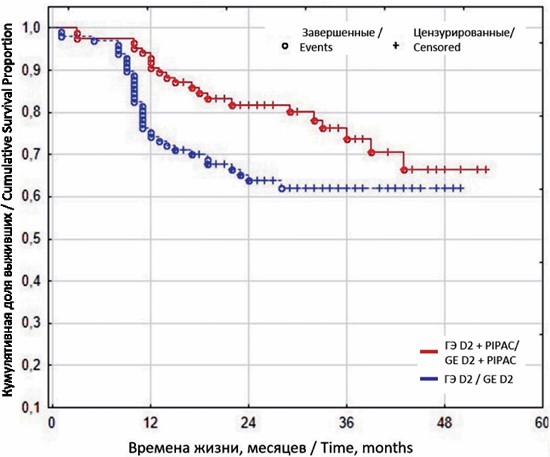
Background. Prevention of peritoneal metastasis remains one of the most critical unresolved challenges in the treatment of locally advanced gastric cancer. The role of prophylactic intraoperative pressurized intraperitoneal aerosol chemotherapy (PIPAC) has not previously been evaluated in a randomized trial. Material and Methods. The prospective randomized GASPACCO trial (NCT04595929) enrolled 182 patients with histologically confirmed gastric adenocarcinoma (cT3a–4bN0–3M0, peritoneal cytology-negative), who completed four cycles of neoadjuvant FLOT chemotherapy. Patients were randomized to receive either radical D2 gastrectomy with a simultaneous intraoperative PIPAC session (cisplatin 7.5 mg/m2, doxorubicin 1.5 mg/m2; n=85) or D2 gastrectomy alone (n=97). All patients received adjuvant FLOT chemotherapy. Follow-up was up to 3 years. overall survival (OS) and recurrence-free survival (RFS) were compared using the log-rank test; rates of peritoneal carcinomatosis were compared using the Pearson chi-square test. Results. PIPAC significantly improved OS at all time points: 1-year OS 94.12 % vs 76.29 % (p=0.0009), 2-year OS 78.12 % vs 61.33 % (p=0.0328), and 3-year OS 78.79 % vs 51.11 % (p=0.0125). Median OS was not reached in either group at the time of analysis. The absolute improvement in OS increased over time: 17.8 % at one year and 27.7 % at three years. the rate of peritoneal carcinomatosis was significantly lower in the PIPAC group: 11.76 % vs 34.02 % (absolute difference 22.26 %, p=0.0004). Mean time to disease progression was significantly longer in the PIPAC group: 26.64 ± 12.95 months vs 21.95 ± 14.46 months (difference 4.69 months, p=0.0175). Conclusion. Prophylactic intraoperative PIPAC added to standard multimodal treatment (neoadjuvant FLOT, radical D2 gastrectomy, adjuvant FLOT) significantly improves overall and recurrence-free survival and reduces the incidence of peritoneal carcinomatosis in patients with locally advanced gastric cancer. These findings support the inclusion of prophylactic PIPAC in clinical treatment protocols for patients at high risk of peritoneal dissemination.

The purpose of the study: to analyze the rationale for routine splenectomy or splenic hilar lymphadenectomy in gastric cancer (GC) surgery through a comparative study of spleen-preserving and splenectomy techniques, and an investigation of the patterns and clinical impact of metastasis of the splenic hilar lymph nodes (SHLN). Material and Methods. A retrospective analysis on data from 264 patients with stage I–III gastric cancer (GC) treated at the Voronezh Regional Scientific and Clinical Oncology Center between 2013 and 2018 was conducted. After examination, all patients underwent total gastrectomy with D2 lymphadenectomy with or without splenectomy (R0 resection). XELOX/FOLFOX adjuvant chemotherapy was utilized if indicated. the median follow-up time was 61 [19–80] months. Results. Metastases to SHLN were found in 19 (7.2 %) patients and were associated with advanced GC (T3–4/N2+) (OR 9.8; 95 % CI 6.76–14.2; p<0.001). At the same time, the techniques used for their removal (splenectomy and spleen-preserving lymphadenectomy) showed limited safety. Despite comparable rates of early postoperative complications between the groups, splenectomy was associated with increased intraoperative blood loss (p<0.001) and a higher incidence of mild pancreatogenic complications (grade II–IIIa according to the Clavien–Dindo classification) (OR 9.67; 95 % CI: 1.24–75.49; p=0.008). In addition, SHLN-status was significantly associated with the development of early postoperative complications following gastrectomy (OR 5.43; 95 % CI 1.59–18.69; p=0.008), as well as with recurrence-free survival (HR 3.21; 95 % CI 1.30–7.95; p=0.01) and overall survival (HR 4.14; 95 % CI 1.88–9.09; p<0.001) in patients with GC. Conclusion. Metastatic involvement of SHLN is associated with locally advanced tumor progression, an unfavorable course of the early postoperative period, and reduced survival in patients with GC. The obtained data emphasize the need for further investigation of a selective approach to lymphadenectomy in this area.

Aim: to evaluate the efficacy of combined neoadjuvant photodynamic therapy with chlorin e6 and proton therapy for uveal melanoma using ultrasound parameters of intratumor blood flow, densitometric, and morphometric indicators. Material and Methods. A retrospective study included 40 patients (40 eyes) with T2–T4 uveal melanoma. All patients were divided into two groups. Group I (n=19) received proton therapy using pencil beam scanning at a total dose of 49 Gy in 7 fractions. Group II (n=21) received photodynamic therapy with chlorin e6 followed by proton therapy. All patients underwent complex ultrasound examination with color doppler imaging and echodensitometry before treatment and during follow-up. Results. Neoadjuvant photodynamic therapy resulted in earlier and more complete tumor devascularization, showing a much higher number of avascular tumors by the 6th month in group II patients (χ2=12.093; p=0.002). Both groups showed gradual tumor regression and decreased acoustic density, but no statistically significant differences in these parameters between the groups were found. During the first 6 months after treatment, combined treatment resulted in a faster drop in tissue acoustic density measured by echodensitometry than proton therapy alone. Conclusion. Neoadjuvant photodynamic therapy combined with proton therapy produces a pronounced vasculo-occlusive effect that peaks by the 6th month. Complex ultrasound examination with color doppler imaging and echodensitometry is an objective tool for assessing tumor regression during organ-preserving treatment.

Objective: to study the morphofunctional state of the recipient zone of adipose tissue transplantation in the lymphadenectomy area after radical breast cancer surgery. Material and Methods. A single-center retrospective-prospective comparative study included 147 patients with histologically confirmed breast cancer, who were treated at Tomsk Regional Oncology Center in 2022–2023. All patients underwent radical mastectomy with axillary lymph node dissection (levels I–III). The study group consisted of 78 patients who underwent axillary lipofilling, and the control group included 69 patients who underwent vacuum drainage. Inclusion criteria: histologically confirmed breast cancer, radical mastectomy with level I–III axillary lymph node dissection, absence of previous surgery or radiotherapy to the axillary region and written informed consent. Exclusion criteria were distant metastases and severe sub-/decompensated comorbidities. The recipient zone was assessed 12–24 months later using imaging techniques and anthropometry of the upper extremities. Ultrasound examination with compression elastometry and shear wave elastography was performed in 21 patients from the study group and in 14 patients from the control group. Lymphoscintigraphy with SPECT/CT was performed in 24 patients from the study group and in 21 from the control group. Results. Comprehensive analysis showed that in 90.5 % of patients in the study group the transplanted adipose tissue was preserved in the recipient zone and had a density corresponding to the reference range of 2–5 kPa. In 19 patients, the median density of the integrated adipose tissue was 3.8 (3.1; 5.9) kPa. The volume of integrated adipose tissue in the recipient zone was 10.50 (7.33; 17.75) cm3 and 11.75 (8.82; 18.50) cm3 after excluding isolated cases of scar replacement. According to compression elastography, the transplanted adipose tissue corresponded to elastotype 2, whereas in the control group the recipient zone demonstrated features of scar tissue. On lymphoscintigraphy of the operated side, the transport index was 13.8 (10.8; 18.8) in the study group and 26.8 (19.3; 32.8) in the control group; background radiopharmaceutical accumulation in the forearm soft tissues was 18 (10; 34) and 42 (28; 66) respectively. Differences between the groups in transport index and background radiopharmaceutical accumulation were statistically significant (p=0.03 and p=0.012). Conclusion. Transplantation of autologous adipose tissue into the axillary lymphadenectomy area ensures its integration in the recipient zone and is associated with more favorable morphofunctional tissue characteristics and lymphatic transport parameters compared with standard drainage.
LABORATORY AND EXPERIMENTAL STUDIES

Objective: to study the expression of alpha-6/beta-4 integrin and laminin-332 in lymph node-positive luminal a breast cancer. Material and Methods. Surgical specimens from 110 patients with luminal a breast cancer (T1–2N0–3M0), who were treated at Tomsk Cancer Research Institute from 2016 to 2022, were analyzed. All patients were divided into two groups: those without lymph node metastases (n=81) and those with lymph node metastases (n=29). The immunohistochemical study was performed according to a standard procedure using antibodies to integrin alpha-6 subunit (ITGA6), integrin beta-4 subunit (ITGB4) and gamma-2 subunit of laminin-332 (LAMG2). Histological samples were digitized using whole-slide scanners. The scanned slices were analyzed using SlideViver V2.8 and QuantCenter software tools. Results. The positive expression of ITGB4 (66 % and 19 %; χ2=22.088; p<0.001) and LAMG2 (86 % and 7 %; χ2=65.514; p<0.001) in tumor cells of the primary tumor was more often detected in patients with lymph nose metastases than in patients without lymph node metastases. Tumor cells positive for ITGA6, ITGB4 and LAMG2 showed higher expression in lymph node metastases than in primary tumors. Conclusion. The expression of alpha-6/beta-4 integrin and laminin-332 correlates with lymph node metastasis, and the expression of gamma-2 subunit of lamnin-332 can serve as a marker for lymph node metastasis.

Introduction. The application of iron and iron oxide nanoparticles for cancer therapy is a promising strategy due to their high surface area, chemical stability, low toxicity and high biological activity against cancer cells. The mechanism of iron oxide-induced toxicity against cells is the production of reactive oxygen species. the investigation of the molecular mechanisms of the effect of particles on cells and the assessment of the influence of the mass ratio of iron to iron oxide on the lipid peroxidation parameters is of great importance for their application. Goals and Objectives: to synthesize core-shell Fe/Fe3O4 nanoparticles to study the effect of iron oxide and iron mass fraction on Hela cell viability and the content of malondialdehyde, bityrosine, catalase, superoxide dismutase and cathepsin D activity and analyze the oxidative modification of proteins in HeLa cancer cell line. Material and Methods. Core-shell Fe/Fe3O4 nanoparticles with an iron oxide mass ratio from 5 to 90 wt. % were synthesized using the electrical explosion of wires in an oxygen-containing atmosphere. Results. The resulting nanomaterials contained a phase composition of Fe, Fe3O4 and FeO, and their biological impact was evaluated by measuring lipid peroxidation levels through standard biochemical assays. The average particle size was approximately 60−80 nm, and the zeta potential of the particles increased from 0.47 to 6.37 mV with increasing mass fraction of iron oxide. The cytotoxicity of the core-shell Fe/Fe3O4 nanoparticles towards HeLa cancer cells was evaluated by an MTT assay and lipid peroxidation parameter determination. The Fe/Fe3O4 nanoparticles with Fe3O4 mass ratio of 40% was found to have a significant effect on the lipid peroxidation intensity, however a maximal protein oxidative modifications accumulation was observed. The correlations obtained need to be taken into account when obtaining iron and iron oxide nanoparticles for cancer treatment. Conclusion. We identified changes in lipid peroxidation and cathepsin D activity in HeLa cancer cell cultures induced by Fe/Fe3O4 nanoparticles. Based on these results, we concluded that Fe/Fe3O4 nanoparticles can be used as an antitumor additive, however their activity depended on Fe3O4 mass ratio. These findings are essential for producing iron and iron oxide nanoparticles for cancer cell targeting.
ONCOLOGY PRACTICE
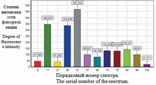
The aim of the study was to improve treatment efficacy for patients with locally advanced nasal cavity and paranasal sinus cancer using intraoperative spectroscopy (IS) and photodynamic therapy (PDT). Material and Methods. To improve immediate and long-term treatment outcomes of combined modality of locally advanced nasal cavity and paranasal sinus cancer. The study included 80 patients with locally advanced nasal cavity and paranasal sinus cancer, who were treated from 2011 to 2024. The patients were divided into two groups: the study group (n=40) and the control group (n=40). Patients in both groups received preoperative radiation therapy at a total dose of 44 Gy followed by surgery. The study group also underwent spectroscopic wound examination and photodynamic therapy during surgery. Results. The five-year relapse-free survival and overall survival rates was significantly higher in the study group (46.0 % and 47 %, respectively) than in the control group (15.0 % and 37.5 %, respectively). Conclusion. The use of intraoperative spectroscopy in combination with photodynamic therapy increase the five-year relapse-free and overall survival rates of patients with locally advanced nasal cavity and paranasal sinus cancer. Photodynamic therapy accelerates wound healing, promotes implant and transplant integration in the recipient zone, and reduces the risk of tumor recurrence.
Background. Immune system is made up of different organs, cells and proteins. a healthy immune system can defeat invading disease-causing germs (or pathogens), such as bacteria, viruses, parasites as well as cancer cells while protecting healthy tissue. White blood cells are the key players in the hematological immune system. Objective. The aim of this research is to detect the radiation therapy effect on the immune system. Material and Methods. Data of 106 cancer patients have been collected from Department of Radiation Oncology, Jinnah Hospital Lahore, Pakistan since January 2018 to September 2022 and compete blood count of these patients is seen regularly before and after therapy. Results. There were 54.2 % female and 45.7 % male patients in the age range of 29–80 years. The 64 % patients were observed with decrease in platelets count, 71.4 % cancer patients had shown a reduction in hemoglobin level and 68.5 % of patients indicated a decline in TLC after the radiation therapy. Conclusion. Conclusions have been drawn from analyzing the data in this research work. Radiation therapy affects the immune system of the patients. Majority of patients have low White Blood Cells Counts after the treatment and also showed low Hemoglobin and Platelets Count.
REVIEWS

Background. Cancer remains one of the leading causes of mortality worldwide. this review analyzes global cancer incidence and mortality rates for 2022 and their association with the Human Development Index (HDI). Material and Methods. The methodology involved a search of the PubMed and eLibrary.ru databases using keywords such as “human potential index”, “cancer incidence worldwide”, “cancer mortality worldwide”, “epidemiology of cancer”, “risk factors affecting cancer”, “Global cancer statistics”. From the relevant articles published between 2019 and 2025, 39 were selected for writing the review. Results. In 2022, there were an estimated 20 million new cancer cases and 9.7 million cancer-related deaths worldwide. The most common cancer sites in terms of incidence and mortality were lung, breast, colon, and liver cancers. among men, cancer of the trachea, bronchi, lung, and prostate predominated; among women, breast cancer was most common. The highest incidence rates especially for gastrointestinal cancers were heavily concentrated in Eastern and Central Asia. The analysis revealed regional disparities. in countries with a high HDI, elevated cancer incidence rates are linked to advanced health care systems and aging population. in low- and medium-HDI countries, the gap between incidence and mortality is small due to the limited access to healthcare, leading to underdiagnosis and low life expectancy. Conclusion. The projected increase in cancer incidence requires healthcare modernization. in high-HDI countries, the increase is driven by a “Western” lifestyle, while in developing countries, the burden of infection-associated cancers remains high. The strategy for reducing the cancer burden should include prevention, universal access to screening and treatment, and monitoring of indicators to assess the effectiveness of measures.
The aim of the study was to summarize current data on the efficacy and safety of BCG immunotherapy for the treatment of patients with non-muscle-invasive bladder cancer. Material and Methods. A literature review was conducted using PubMed, Cochrane Library, and E-library databases, primarily selecting sources from the last 10 years. Key words used in the database search included bladder cancer, BCG immunotherapy, BCG therapy safety, BCG therapy efficacy, non-muscle-invasive bladder cancer, NMIBC, SWOG, and adjuvant therapy in oncology. in preparation for writing the literature review, 39 articles from the 148 retrieved articles were selected. Results. This literature review summarizes domestic and international data on the history of the development and implementation, efficacy, and safety of BCG immunotherapy in the treatment of bladder cancer. The limitations of use and challenges of modern healthcare in implementing adjuvant BCG therapy for cancer patients were also discussed. a separate aspect of the review was the safety and algorithms for preventing and treating specific complications of BCG immunotherapy (granulomatous lesions of the urinary tract, BCG sepsis). Conclusion. Intravesical BCG immunotherapy in the treatment of patients with non-muscle-invasive bladder cancer at intermediate and high risk of recurrence is the method of choice for adjuvant therapy. The review showed that in real-world clinical practice, BCG therapy is used to treat a much smaller proportion of patients than indicated. Moreover, the administrative, legal, and clinical restrictions that specialists fear are often easily surmountable. In turn, high-quality TB care during the selection stage for therapy and for follow-up helps identify patients with contraindications to immunotherapy and promptly diagnose tuberculosis. However, the necessity, effectiveness, and safety of prophylactic treatment for tuberculosis before BCG immunotherapy remain unclear and requires further study.
Objective: to summarize current trends in the use of abiraterone in the treatment of prostate cancer based on clinical and experimental studies. Material and Methods. We searched and analyzed information in MedLine, Scopus, WoS, and the Russian Science Citation Index (RSCI) databases for the past 10 years. We analyzed information on pharmaceutical, experimental, and clinical studies on current trends in abiraterone use strategies for prostate cancer treatment, based on clinical and experimental studies. Results. Abiraterone-based therapies are being actively studied for the treatment of both hormone-sensitive and castration-resistant prostate cancer. The use of abiraterone-based therapies or irreversible androgen receptor blockers leads to long-term disease control and improved treatment outcomes for prostate cancer patients. Regulation of testosterone levels between castration and supraphysiological levels suppresses the growth of cancer cells (bipolar androgen therapy). Significant limitations in the use of abiraterone-based therapies are associated with their side effects: changes in water-electrolyte balance due to the effect on the level of mineralocorticoids in the blood, and the development of relative or absolute liver and kidney failure. To reduce the dose and/or increase the bioavailability of the drug, two main approaches are used: using the positive effect of food to increase exposure and the development of supergenerics. Current strategies for overcoming resistance to deprivation and chemotherapy include combination therapy using agents with different mechanisms of pharmacological activity, metronomic administration, and the development of new modified antitumor agents that combine a cytotoxic and antiandrogenic component in a single molecule. Pharmacologically active compounds, such as abiraterone, have been developed using organotin compounds, offering dual (multimodal) action and actively inducing apoptosis and inhibiting proliferation in the G2/M phase of the cell cycle (in vitro studies). Conclusion. The combination of these approaches has the potential to be used for personalized treatment of hormonally active malignant tumors in various locations (prostate, breast, etc.).

Purpose of the study. The aim of this review was to analyze the role of clinical registries, prognostic models, and decision support systems in the surgical treatment of colorectal cancer, and to substantiate their relevance for preventing postoperative complications and improving short-term treatment outcomes. Material and Methods. The review highlights the current scientific literature, clinical guidelines, population-based studies, national quality audits, cancer registries, and publications addressing the prediction of surgical outcomes, artificial intelligence, and machine learning in colorectal surgery. Particular attention was paid to studies analyzing postoperative complications, inter-center variability in outcomes, death after severe postoperative complications, composite indicators of surgical quality, and the potential clinical application of prognostic tools. Results. Colorectal cancer remains a major epidemiological burden and requires further improvement in the quality of surgical care. Postoperative complications after colorectal cancer resections occur in a substantial proportion of patients and are associated with longer hospital stay, higher rates of repeat interventions, early mortality, and a potential adverse effect on long-term prognosis. Minimally invasive techniques and enhanced recovery protocols contribute to improved short-term outcomes; however, their effectiveness depends on institutional experience, the organization of perioperative care, and the ability of the healthcare system to identify and manage complications early, thereby preventing failure-to-rescue. Clinical registries provide an opportunity for population-level assessment of treatment outcomes, identification of inter-center variability, analysis of factors associated with adverse outcomes, and development of datasets for prognostic models. modern predictive tools, including artificial intelligence and machine learning methods, have potential for risk stratification but require external and prospective validation, clinical interpretability, and integration into real-world clinical workflows. Conclusion. Registry-based decision support systems may be considered a tool for shifting from retrospective outcome analysis to proactive quality management in surgical care for colorectal cancer. Their practical value is determined not only by the accuracy of prognostic models, but also by their ability to translate risk assessment into clinical actions, patient pathway management, individualized perioperative care, and early escalation of treatment.
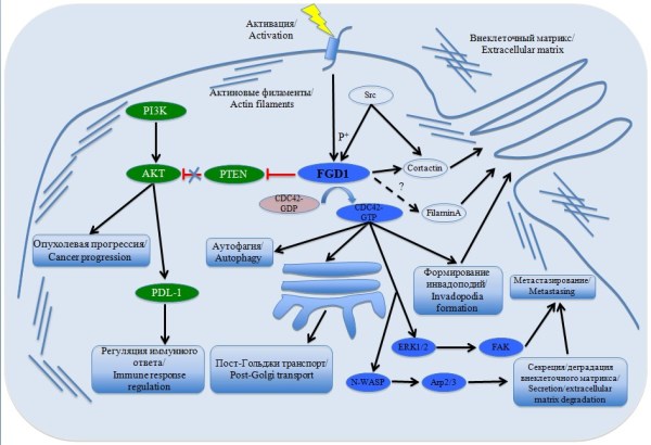
The aim of the study was to systematize and summarize current data on the role of the faciogenital dysplasia 1 protein (FGD1) in the mechanisms of progression and metastasis of multiple malignancies including breast carcinoma, osteosarcoma, melanoma, hepatocellular carcinoma and acinar prostate carcinoma. Material and Methods. We reviewed local and international publications registered in the MedLine/ PubMed databases over the past 15 years. Results. The review examines the molecular mechanisms by which FGD1 regulates cancer progression and metastasis. The FGD1 gene, located at the X chromosome, encodes a guanine nucleotide exchange factor (GEF) that specifically activates the gtpase CDC42. Through its effectors, FGD1 is involved in numerous signaling pathways, including gene expression, membrane transport, cell polarization, and cytoskeleton organization. Conclusion. A literature review revealed that the FGD1 protein might be a promising target molecule. Further studies of the interactions between the FGD1/CDC42 and PI3K/AKT signaling pathways will expand treatment options for multiple malignancies
CASE REPORTS

Background. Gastric neuroendocrine tumors (gNETs) are rare neoplasms accounting for less than 3 % of all gastric tumors. Type 1 gNETs are the most common tumors associated with autoimmune gastritis and characterized by a low risk of metastasis and a favorable prognosis. Case description. We report a rare case of regional metastasis of multiple type I gNETs in a 35-year-old male patient with a history of autoimmune atrophic gastritis. Endoscopic ultrasonography revealed more than 20 subepithelial lesions measuring up to 1.5 cm in the gastric fundus and body, with the largest involving the submucosal layer. Large tumors were excised using endoscopic submucosal dissection (ESD). Histological and immunohistochemical studies revealed well-differentiated gNETs G1 (Ki67 – up to 2 %) with invasion into the submucosal layer and signs of incomplete removal of the tumor (pT1b L1V0R1). The patient underwent laparoscopic gastrectomy with D2 lymph node dissection. Histological examination of the surgical specimen revealed subcapsular metastasis of gNET in one of the removed lymph nodes with invasion of its capsule, but no tumor cell spread beyond the node. The patient did not receive any specific therapy pre- or postoperatively. Conclusion. This clinical case demonstrates that even with a favorable immunophenotype of gNETs type I, a tumor size greater than 1.0 cm and invasion into the submucosal layer indicate an increased risk of lymph node metastasis. This case confirms the validity of current clinical guidelines that consider these factors as key criteria for making decisions on surgical treatment with lymph node dissection.

Objective: to present a clinical case of delayed dual-tracer sentinel lymph node biopsy using technetium-99m radiocolloid and indocyanine green fluorescence in a patient who previously underwent nipple-sparing mastectomy with immediate implant-based breast reconstruction, and to analyze the technical feasibility and early perioperative safety of the procedure. Material and Methods. This article presents a clinical case of delayed sentinel lymph node biopsy after previous nipple-sparing mastectomy with immediate implant-based breast reconstruction. The report is descriptive and was not intended to assess diagnostic accuracy, sensitivity, specificity, or the false-negative rate of the technique. A 40-year-old woman with a history of right nipple-sparing mastectomy and immediate implant-based reconstruction underwent delayed axillary sentinel lymph node biopsy for staging regional lymphatic basin. Preoperative lymphatic mapping was performed using periareolar subcutaneous injection of 50 MBq 99mTc-Nanocoll, followed by lymphoscintigraphy and SPECT-CT. On the day of surgery, indocyanine green was injected intradermally at four periareolar points. intraoperative detection was performed using a near-infrared fluorescence imaging system, MARS, version 2.0.0.7, 2024, and a handheld gamma probe. The primary procedural endpoint was technical success, defined as intraoperative identification and excision of at least one sentinel lymph node. Results. Preoperative lymphoscintigraphy and SPECT-CT demonstrated radiotracer uptake in two right axillary lymph nodes. Intraoperatively, both sentinel lymph nodes were successfully identified and excised using combined gamma-probe detection and fluorescence guidance. Concordance between radioactive and fluorescent signals was observed in both removed nodes. Histological examination showed no metastatic involvement. No intraoperative or early postoperative complications were recorded. The implant, mastectomy flaps and nipple-areolar complex remained viable, with no evidence of ischemia, infection, implant exposure or reconstructive failure. Conclusion. In this clinical case, delayed dual-tracer sentinel lymph node biopsy was technically successful and was not associated with early perioperative complications. These findings indicate the principal technical feasibility of the procedure in this specific clinical situation, but they do not allow assessment of diagnostic accuracy, reproducibility, or oncological safety.
ANNIVERSANES


ISSN 2312-3168 (Online)