

LABORATORY AND EXPERIMENTAL STUDIES
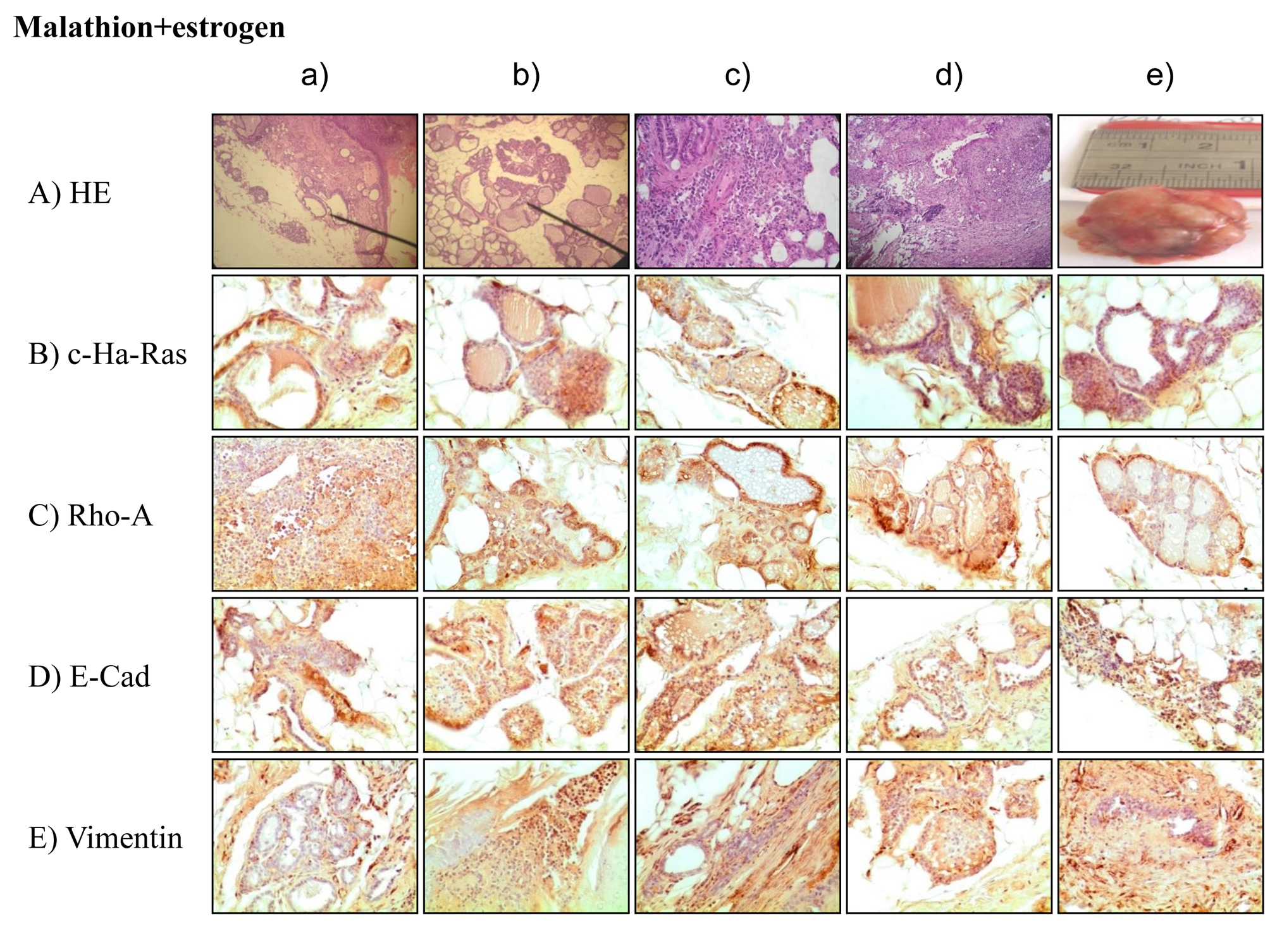
Breast cancer is considered a major and common health problem in both developing and developed countries. The etiology of breast cancer, the most frequent malignancy diagnosed in women in the western world, has remained unidentified. Chemicals as the organophosphorous pesticide malathion have been used to control a wide range of sucking and chewing pests of field crops, and are involved in the etiology of breast cancers. The association between breast cancer initiation and prolonged exposure to estrogen suggests that this hormone may also have an etiologic role in such a process. However, the key factors behind the initiation of breast cancer remain to be elucidated. The effect of environmental substances, such as malathion and estrogen was analyzed in an experimental rat mammary gland model. Different cytoplasmic proteins are key in the transformation of a normal cell to a malignant tumor cell and among these are the Ras super family and Ras homologous A (Rho-A). Both types of proteins were greater in animals treated with malathion than those with estrogens. E-Cadherins constitute a large family of cell surface proteins.
Results showed greater expression of E-Cadherin and vimentin than c-Ha-ras and Rho-A in rats treated by estrogens. In breast cancer, analysis using immunohistochemical markers is an essential component of routine pathological examinations, and plays an important role in the management of the disease by providing diagnostic and prognostic strategies.
The aim of the present study was to identify markers that can be used as a prognostic tool for breast cancer patients.
Dendritic cells (DCs) belong to specialized pool of antigen-presenting cells with high functional plasticity and manifest with immunostimulatory or immunosuppressive potential depending on sequence and combination of microenvironment stimuli, which determine their differentiation, maturation and activation. The use of antitumor DCs vaccines is based on the ability of DCs specifically activated in vitro migrate for antigen presentation to T- lymphocytes. We studied the components of the tumor microenvironment that are capable of inhibiting DCs migration. The study of the mobility of DCs in Cell-IQ experimental analytical system showed the presence of an inverse correlation of high strength between the average trajectory speed and the level of immunosuppressive factors (ISFs) in supernatants of cultured skin melanoma cells (TGFβ1, IL-10, IL-18, VEGF-A, EGF, FGF, HGF, sFASL (p<0.01). An inverse relation of high force was found between the movement angle of the DCs population and the expression of cancer testis antigens (CTAs) and other tumor- associated antigens (TAAs) on tumor cells (Melan A, tyrosinase, families of MAGE, BAGE, NY- ESO-1 (p<0.01)). The speed of DCs movement in culture system with melanoma cells #894 was 30.10±2.23 μm/h and differed from that in the presence of IL-10 1 ng/ml (10.45±0.52 μm/h), TGFβ1 10 ng/ml (14.32±0.42 μm/h), VEGF 50 ng/ml (18.7±1.89 μm/h) (p<0.05). One can assume that content of this ISFs in the blood is one of the factors determining clinical efficacy of DCs immune therapy.
Using DIC and confocal microscopy, changes in morphology, migratory characteristics and adherence junctions (AJs) were analyzed in the mammary carcinoma cell line MCF-7-SNAI1 after activation of the EMT transcription factor SNAI1. Western Blot analysis showed that after removal of tetracycline from the cell culture medium expression of SNAI1 reached its peak in 24 hours and then plateaued for 7 days. During the 7 days the cells continued to express E-cadherin; however, tangential AJs typical for cells with stable cell-cell adhesion, changed into radial AJs. The radial AJs continued to accumulate E-cadherin during 24‑72 hours after tetracycline removal. As a result of SNAI1 activation, the cells underwent epithelial-mesenchymal transition (EMT) and became migratory. On a two-dimensional substrate, cells exhibited both individual and collective migration. As the tetracycline washout period progressed, the fraction of the cells capable of migrating through migration chamber membranes increased; on the contrary, cells’ ability to invade an epithelial monolayer decreased. These results demonstrate that retaining a hybrid epithelial/mesenchymal phenotype and accumulation of E-cadherin in AJs during early stages of EMT do not impede disruption of stable cell-cell adhesion and cells’ acquisition of migratory activity.
PCR clamping/wild-type blocking PCR with non-extendable locked nucleic acid (LNA) oligonucleotides is used for sensitive detection of somatic mutations in tumors. Various versions of the technique use different DNA polymerases and LNA oligonucleotides with and without additional phosphorothioate modifications. Here we studied requirements for successful PCR clamping with LNA oligonucleotides and Taq DNA polymerase for analysis of mutations in KRAS and BRAF genes by means of real-time PCR and Sanger sequencing. We found that addition of phosphorothioate linkages at the 5’-end of LNA oligonucleotide to protect from 5’- exonuclease activity of Taq DNA polymerase did not improve clamping. For most target sequences, efficient clamping was observed at melting temperature of LNA oligonucleotide 20‑25°C above annealing/extension temperature of the PCR with a 2-step protocol. Under such conditions, simple and sensitive detection of mutations in KRAS and BRAF genes was feasible using real-time PCR with TaqMan probes or Sanger sequencing.
The exosomes involvement in the pathogenesis of tumors is based on their property to incorporate into the recipient cells resulting in the both genomic and epigenomic changes. Earlier we have shown that exosomes from different types of estrogen-independent breast cancer cells (MCF-7/T developed by long-term tamoxifen treatment, and MCF-7/M) developed by metformin treatment were able to transfer resistance to the parent MCF-7 cells. To elucidate the common features of the both types of resistant exosomes, the proteome and microRNA cargo of the control and both types of the resistant exosomes were analyzed. Totally, more than 400 proteins were identified in the exosome samples. Of these proteins, only two proteins, DMBT1 (Deleted in Malignant Brain Tumors 1) and THBS1 (Thrombospondin-1), were commonly expressed in the both resistant exosomes (less than 5% from total DEPs) demonstrating the unique protein composition of each type of the resistant exosomes. The comparative analysis of the miRNA differentially expressed in the both MCF-7/T and MCF-7/M resistant exosomes revealed 180 up-regulated and 202 down-regulated miRNAs. Among them, 4 up-regulated and 8 down-regulated miRNAs were associated with progression of hormonal resistance of breast tumors. The bioinformatical analysis of 4 up-regulated exosomal miRNAs revealed 2 miRNAs, mir- 101and mir-181b, which up-regulated PI3K signaling supporting the key role of PI3K/Akt in the development of the resistant phenotype of breast cancer cells.
CLINICAL STUDIES
Currently the impact of autophagy on carcinogenesis remains understudied. On the one hand, autophagy acts as a tumor suppressor, as it activates degradation of oncoproteins, toxic proteins, and damaged cell organelles, that may be aggressive and lead to DNA damage. On the other hand, autophagy may promote tumor cell survival under hypoxia and in the presence of reactive oxygen species, which occurs primarily due to blocking of apoptosis mechanisms, raising the chances for maintaining tumor clone dynamics. Autophagy regulation is a complicated and multi-stage process. The main regulator here is a signaling pathway that activates serine/threonine protein kinase m-TOR (the mammalian target of rapamycin). Data on the impact of autophagic proteins ATG5, LC3A, LC3B, and Beclin-1 on malignant cell survival as well as on tumor growth and progression have been reported in literature. However, studies aimed at seeking possible relationships between autophagy and pathogenetic mechanisms of carcinogenesis are of great interest.
The aim of the study is to investigate a relationship between the expression parameters of autophagy regulatory proteins m-TOR and Beclin-1 and the features of lymphogenic metastasis in colorectal cancer.
Materials and methods. The study included 105 patients with T1-4N0-3M0 colorectal cancer treated in the Thoracic and Abdominal Department of Cancer Research Institute of Tomsk Research Medical Center from 2012 to 2015. The average age of patients was 59.7±4.3 years. Morphological verification of the diagnosis was performed on the biopsy samples of primary tumor tissue. Staging of colorectal cancer was determined according to the TNM classification of malignant tumors (2002).
Results. Analysis of the frequency of lymphogenic metastasis depending on the presence or absence of m-Tor and Beclin-1 expression in tumor cell cytoplasm revealed a statistically significant link between these variables.
Conclusion. The obtained findings clearly exhibit that deceleration or loss of autophagic activity in the tumor is accompanied by implementation of lymphogenic dissemination, which is a predictor of an unfavorable outcome of the disease.
For patients with an identified germline E-cadherin-1 (CDH1) mutation, prophylactic gastrectomy is the treatment of choice to eliminate the high risk of developing diffuse gastric cancer. The case report describes a rare case of hereditary diffuse gastric cancer (HDGC) associated with CDH1 gene mutation, which is reported in the Russian population for the first time. In 2013, a 28-year- old woman was referred to Clinical Oncogenetics Laboratory with a family history of gastric cancer. Molecular genetic analysis revealed CDH1 gene mutation. The lifetime risk of cancer in mutation positive members is more than 80. Histological examination of gastric biopsy specimens obtained during endoscopy revealed isolated signet ring cells in the lamina propria. Spleenpreserving D2-lymphodissection and total gastrectomy with Roux-en-Y reconstruction with a jejunal reservoir formation were performed at the Abdominal Oncology Surgery Department.
Aim of the study. Aim of the study was to estimate the occurrence of pathogenic mutations in the BRCA1 gene in Russian breast cancer patients.
Material and methods. Complete coding sequence of the BRCA1 gene of 445 early onset breast cancer patients (under 40 years) from Novosibirsk region (Russia) were analyzed by targeted Next Generation Sequencing (NGS) using Ion Torrent platform. Results. Forty (9%) carriers of various pathogenic mutations were revealed. Thirty five (7,9%) patients carried 5382insC mutation, described earlier as a founder mutation for Slavic population. Five (1.1%) patients carried various pathogenic mutations, namely C61G, 462delCC, E143X, 4153delA, and IVS18+1G>T. Besides, 29 genetic variants with no clinical significance or with unknown clinical significance were detected in BRCA1 gene among 445 early onset breast cancer patients. Conclusions. Data on the frequency of genetic variations in the BRCA1 gene among early onset breast cancer patients in the Novosibirsk Region (Russia) were obtained. Proportion of the 5382insC mutation is 87.5% of all pathogenic mutations in the BRCA1 gene found in patients.
Introduction. Intratumor heterogeneity is a characteristic feature for most malignant tumors, including cutaneous melanoma. This property represents one of the main obstacles for effective targeted therapy, due to the different sensitivity to chemotherapeutic agents on various tumor cells subclones. Treatment of malignant tumors requires an individual approach to choose the most appropriate treatment regimen.
The purpose of the study was to evaluate differences in melanoma tissue samples obtained from different parts of one patient’s primary tumor at the transcriptomic level.
Material and Methods. Melanoma cell cultures obtained from both central and peripheral parts of the primary tumor of two patients were used in the study.
Results. Subclones from different parts of the first patient’s tumor were similar, whereas the second patient demonstrated significant differences at the transcriptomic level (in 2953 transcripts out of 48226). In the cells of the central zone of the second patient’s tumor, an increase in mRNA of the genes encoding proteins associated with tumor-specific immune response, as well as ABC-family transport proteins and cytokine signaling molecules, were noted. In the cells from the peripheral area of the same tumor, a more intensive transcription of genes encoding extracellular matrix and inflammatory response proteins was observed. Taken all round, the differences between the subclones of the second patient’s cells were relevant to some signaling cascades playing a leading role in oncogenesis (MAPK, PI3K-Akt-mTOR, VEGFA-VEGFR2, etc.).
Conclusion. The study allowed evaluation of differences between cancer cells within a tumor at the transcriptional level in order to search for further approaches to personalized melanoma therapy.
Introduction. The efficacy of anticancer treatment depends on biological factors of tumor.
The aim of the study was to determine the activity of proteasomes and calpains and to reveal their association with VEGF, HIF-1α and NF-κΒ expressions in normal, primary and metastatic renal cell carcinoma (RCC) tissues.
Methods. Ninety-three patients with renal cell carcinoma were included into the study. The expression levels of transcription factor and VEGF were measured using ELISA kits. The levels of proteasome subunits were measured by Western Blotting. Proteasome and calpain activities were determined using specific fluorogenic substrates.
Results. We revealed inactivation of proteolysis in patients with kidney cancer. Disease advance was associated with a significant depression of cellular proteolysis and increase in transcription and growth factor levels in primary kidney cancer tissues. The proteolysis activation was found in metastatic tissues.
Conclusions. Our results suggest that NF-κΒ, HIF-1α and VEGF transcription factors and intracellular proteolytic systems are involved in kidney cancer progression.
The aim of this study was to assess CXCR4 expression in different subsets of CTCs and single (detached) breast cancer cells.
Materials and methods. Thirty five patients with invasive breast carcinoma of no special
type (IC NST) (T1-4N0-2M0), between 29 and 69 years of age were included in this study. Different subsets of CTCs with CXCR4 expression were evaluated by flow cytometry. A confocal microscopy was used to assess CXCR4 expression in different subsets of single (detached) cancer cells in breast tissue.
Results. The CXCR4 was expressed in CTCs without stem-like and EMT phenotype, in CTCs with EMT but not stem markers and in stem-like CTCs without EMT features. In all blood samples, the CXCR4 expression in CTCs with stem-like and EMT phenotype was absent. In breast tumor the CXCR4 was expressed in the non stemlike single (detached) breast cancer cells with EMT features, in the single (detached) breast cancer cells with stem and EMT features. In all tumor samples the stem-like or non stem-like single (detached) breast cancer cells without EMT features were absent.
Conclusions. Different subsets of the CTCs exhibited CXCR4. The CXCR4 expression did not depend on the presence or absence of stem or/and EMT features in tumor cells. We showed that some subsets of single (detached) breast cancer cells in the primary tumor were characterized by the ability to express CXCR4 and may be a source of the respective CTC subsets.
Introduction. The purpose of this study was to evaluate the feasibility of using 99mTc-TG SPECT in the detection and staging of malignant lymphoma.
Materials and methods. Fifteen patients with newly diagnosed malignant lymphoma underwent 99mTc-TG SPECT. Six patients had Hodgkin’s lymphoma and 9 patients had aggressive forms of non-Hodgkin’s lymphoma (NHL): diffuse large B-cell lymphoma (7 cases), B-cell follicular lymphoma (1 case), and lymphoma from B cells in the marginal zone (1 case). Stage IIA was diagnosed in 5 patients, stage IIB in 1, stage IIIA in 1, stage IVA in 4 and stage IVB in 4 patients.
Results. Pathological 99mTc-TG uptake in lymph nodes was observed in 14 (93 %) of the 15 patients. In one patient, the enlarged submandibular lymph node (16 mm in size) detected by CT was not visualized by 99mTc-TG SPECT. This false-negative result was likely to be associated with increased accumulation of 99mTc-TG in the oropharyngeal region. There were difficulties in the visualization of paratracheal, para-aortic and paracardial lymph nodes. These difficulties were associated with a high blood background activity, which persisted even 4 hours after intravenous injection of 99mTc-TG. Software-based SPECT and CT image fusion allowed visualization of these lymph nodes. The pathological 99mTc-TG accumulation in axillary, supraclavicular, infraclavicular and cervical lymph nodes was observed most often. Extranodal involvement was seen in 9 patients. 99mTc-TG SPECT identified extranodal hypermetabolic lesions in 7 (78 %) of these patients. In one patient, hypermetabolic lesion in the lung detected by 99mTc-TG SPECT was not detected on CT image. CT identified bone marrow involvement in the pelvic and scapula in 1 patient. The use of 99mTc-TG SPECT allowed the visualization of hypermetabolic bone tissue lesions in this patient (Figure 4). In addition, in a patient with intact bone tissue on CT, 99mTc-TG SPECT detected hypermetabolic lesions in the iliac bone.
Conclusion. 99mTc-1-Thio-D-glucose demonstrated increased uptake in nodal and extranodal sites of lymphoma. The results indicate that SPECT with 99mTc-1-Thio-D-glucose is a feasible and useful tool in the detection and staging malignant lymphoma.
REVIEWS
Introduction. Colon and rectal cancer (CC, RC) are different entities from a clinical and tumor biological point of view. Up to now, both, CC and RC, are synonymously called “Colorectal Cancer” (CRC). With our experience in basic and clinical research and routine work in this field we now have come to the opinion, that the term “CRC” should definitely be questioned, and if justified, be abandoned.
Materials/Methods. We analyzed the actual available data from the literature and our own results from the Ulm based study group FOGT to proof or reject our hypothesis.
Results. The following evident differences were recognized: Anatomically, the risk to develop RC is 4× higher than for CC. Molecular changes in carcinogenesis in CC are different from RC. Physical activity helps to prevent CC, not RC. Pathologically there are differences between RC and CC. In addition, there are also major clinical differences between CC and RC, such as in surgical topography and– procedures, multimodal treatment (MMT) approaches (RC in MMT is less sensitive to chemotherapy than CC), and prognostic factors for the spontaneous course and for success of MMT (e.g. TS or DPD ). Discussion. CC ´sand RC´s definitely are different in parameters of causal and formal carcinogenesis, effectivity of primary prevention by physical activity, conventional and molecular pathology.According to our findings we can demand from the preclinical point of view that CC and RC are two different tumor entities in terms of various representative biological characteristics.CC and RC are also differing substantially in many clinical features, as outlined in a separate paper from our group.
Conclusion. “CRC” should no longer be used in basic and clinical research and other fields of cancer classification as a single disease entity. CC is not the same as RC. CC might even be divided into right and left CC.
In the present review we collected the main studies regarding the role of chitinase-like proteins (CLPs), belonging to the family of Glyco_18 domain-containing proteins, in different cancers. In humans, 3 chitinaselike proteins have been identified: YKL-40 (CHI3L1), YKL-39 (CHI3L2) and stabilin-1-interacting chitinase-like protein (SI-CLP). CLPs are produced by several types of cells and combine the properties of cytokines and growth factors. The high levels of CLPs were identified in the circulation of the patients with inflammatory diseases and various types of tumors. We highlighted the main known functions of CLPs in normal and pathological conditions, their contribution to metastasis development, angiogenesis, invasion and other processes in cancer, the correlation of the levels of CLPs with tumour progression. Our data also contribute to the understanding of question how CLP could be useful for cancer patient benefit.

ISSN 2312-3168 (Online)


