CLINICAL STUDIES
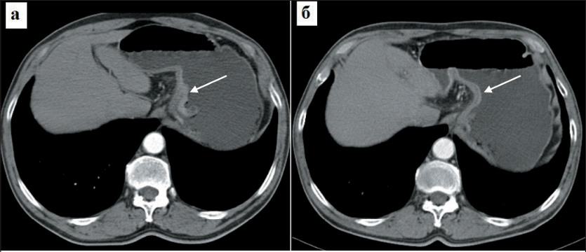
Background. Gastric cancer remains one of the leading causes of cancer-related mortality globally and ranks second in Russia. Pathological complete response (pCR, ypT0N0M0) is a significant surrogate marker for the efficacy of neoadjuvant therapy and disease prognosis. Despite accumulating data, the demographic and clinical-morphological characteristics of this patient category, as well as the dependence of pCR on the neoadjuvant therapy regimen, remain insufficiently studied.
Aim: to characterize the demographic and clinical-morphological characteristics, methods and tolerability of perioperative therapy, and features of surgical treatment in a multicenter retrospective cohort of patients with locally advanced gastric and esophagogastric junction cancer, who achieved pCR after neoadjuvant therapy.
Material and Methods. Data from 221 patients with histologically confirmed gastric or esophagogastric junction adenocarcinoma (cT2-4N0-3M0) who achieved pCR (ypT0N0M0) confirmed by pathomorphological examination of the resected specimen were retrospectively analyzed.
Results. The median age was 65 years (range 24-89). Men (52.5 %) and patients with clinical stage iii (48.0 %) and IIB (30.8 %) predominated. Poorly differentiated adenocarcinoma was detected in 124 (56.1 %) patients, and signet ring cell carcinoma in 35 (15.8 %). Lymph node metastasis (cN+) at the time of diagnosis was found in 124 (56.1 %) patients. The completion rate of neoadjuvant chemotherapy was 96.6 %. Grade >3 adverse events were registered in 17 (8.3 %) patients receiving chemotherapy and in 3 (17.6 %) receiving chemoradiotherapy. According to follow-up examination, a complete clinical response was detected in 2 (0.9 %) patients, a partial response in 57 (25.8 %), and disease stabilization in 162 (73.3 %). Adjuvant chemotherapy was administered to 132 (59.7 %) patients (including the FLOT regimen in 75.0 %). The predominant method of surgical treatment was gastrectomy (59.3 %). Postoperative complications occurred in 47 (21.3 %) patients, including Clavien-Dindo grade >iii in 24 (10.9 %); the 30-day postoperative mortality rate was 1.4 %.
Conclusion. Within the clinical study, the largest sample of the Russian population of gastric cancer patients with a registered pathological complete response after neoadjuvant therapy was collected - 221 patients. This cohort is dominated by features associated with aggressive gastric cancer: diffuse and mixed Lauren types; poorly differentiated adenocarcinoma, including signet ring cell carcinoma; clinical categories cT3-4 and cN+. in patients with pCR, the completion rate of neoadjuvant therapy was 96.6 %, with the FLOT regimen being the most frequently used. Routine CT scanning does not reliably diagnose pCR; thus, searching for and validating new standardized biomarkers for the objective assessment of tumor response to neoadjuvant therapy is highly relevant.
The purpose of the study was to improve radiotherapy efficacy by optimizing fractionation regimens in patients with inoperable stage llb-lll locally advanced pancreatic cancer.
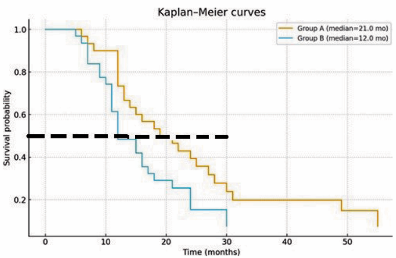
Material and Methods. Treatment outcomes of 60 patients with inoperable stage llb-lll locally advanced pancreatic cancer were analyzed in a single-center comparative study with a retrospective analysis of prospectively collected clinical data. All patients received chemoradiotherapy, including 3 or more cycles of polychemotherapy (PCT) with FOLFlRlNOX or GEMCAP regimen in combination with daily split-dose radiation therapy (RT) (group A) or stereotactic radiotherapy (STRT) (group B).
Results. The efficacy of daily split-dose RT was found to be higher than that of stereotactic RT Early radiation reactions corresponded to grade l-ll and were observed in both groups. lncreased levels of transaminases were the most common: in 36.7 % of patients in group A and in 46.7 % in group B (p=0.59). Nausea/vomiting was observed in 23.3 % and 40.0 %, respectively (p=0.18). An analysis of the immediate results showed that the overall rate of objective response was significantly higher in group A compared with group B (93.3 vs 70.0 %; p=0.03). However, disease progression was observed more frequently in group B than in group A patients (30.0 vs 6.7 %; p=0.042). The daily split-course regimen demonstrated significantly higher one-year (73 vs 48 %) and two-year (39 vs 15 %) overall survival rates than stereotactic RT (p<0.05).
Conclusion. Our study demonstrated the use of combination therapy in a carefully selected group of patients with inoperable stage llb-lll locally advanced pancreatic cancer. This type of therapy allows for increased life expectancy, overall survival, and objective response rates. Moreover, continuous improvements in radiation therapy techniques help reduce the incidence of early systemic radiation reactions. Thus, the daily split-dose dose RT is more effective and can be considered the preferred RT regimen for the treatment of inoperable patients with locally advanced pancreatic cancer.
Objective: to evaluate the efficacy and safety of sequential treatment with radium-223 chloride (Ra-223) followed by 177Lu-PSMA in patients with metastatic castration-resistant prostate cancer (mCRPC) in real-world clinical practice.
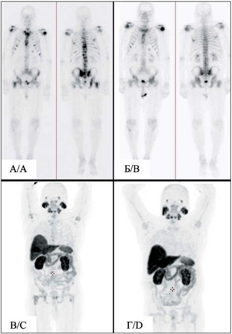
Material and Methods. This retrospective single-center study included 16 mCRPC patients who received at least 4 Ra-223 injections followed by 177Lu-PSMA therapy (at least 2 cycles) between 2022 and 2026. The median age was 69.5 years. All patients had bone metastases; 94 % had prior docetaxel treatment. We assessed biochemical response (prostate-specific antigen (PSA) decline >50 %), radiological response using PSMA PET/CT and bone scintigraphy, and toxicity according to CTCAE v5.0.
Results. Twelve patients received a full course of Ra-223 (6 injections). Biochemical response to Ra-223 was observed in 2 (12.5 %) patients, radiological response in 5 (31.3 %). After a median interval of 2.2 months, 177Lu-PSMA therapy was initiated (median 5 cycles). Biochemical response to 177Lu-PSMA was achieved in 11 (68.8 %) patients, including 7 out of 9 who had progressed on Ra-223. Radiological response was recorded in 9 (56.3 %) patients. Median PSA level decreased from 130.5 to 15.9 ng/mL. Severe (grade 3) hematological toxicity occurred in 2 (12.5 %) patients, xerostomia in 3 (18.8 %).
Conclusion. Sequential therapy with Ra-223 and 177Lu-PSMA demonstrates high efficacy (68.8 % biochemical response rate) and an acceptable safety profile in mCRPC patients, including those with progression on Ra-223, supporting the feasibility of this therapeutic strategy in clinical practice.
Objective: to assess the quality of life of patients with breast and reproductive system cancers, as well as to study the emotional response of these patients and evaluate the level of anxiety, depression and attitudes towards death.
Material and Methods. The study was conducted at the A. Tsyb Medical Radiological Research Centre - branch of the National Medical Research Radiological Centre of the Ministry of Health of the Russian Federation (Obninsk), and the Department of Gynecological Oncology and Breast Tumors No. 2 (Moscow). A total of 118 women diagnosed with breast cancer and 102 healthy women with breast implants for aesthetic purposes were examined. A clinical and psychopathological examination combined with psychodiagnostic testing were performed.
Results. A correlation analysis using Spearman's rank correlation criterion revealed a positive correlation between pain intensity and social functioning at a trend level of 0.7. A similar trend was found in the direct correlation between the general health scale and the mental health and social functioning scale - 0.46 and 0.57, respectively. An inverse correlation was also found between the general health scale and anxiety level scores (R=-0.51). Conclusions. Breast cancer patients exhibit high levels of state anxiety and moderate depression. However, thoughts of death frequently become a fact of life that patients eventually process to find a sense of relief and acceptance.
LABORATORY AND EXPERIMENTAL STUDIES
Background. The treatment efficacy of stage I-II cervical cancer (CC) remains suboptimal due to the risk of recurrence and metastasis, which complicates the selection of optimal treatment strategies and necessitates the search for prognostic markers.
This study aimed to identify molecular characteristics and markers associated with progressive stage I-II CC.
Material and Methods. Clinical and morphological analyses and whole-transcriptome sequencing were performed, and publicly available data from the Cancer Genome Atlas (TCGA) database were used to validate the sequencing results.
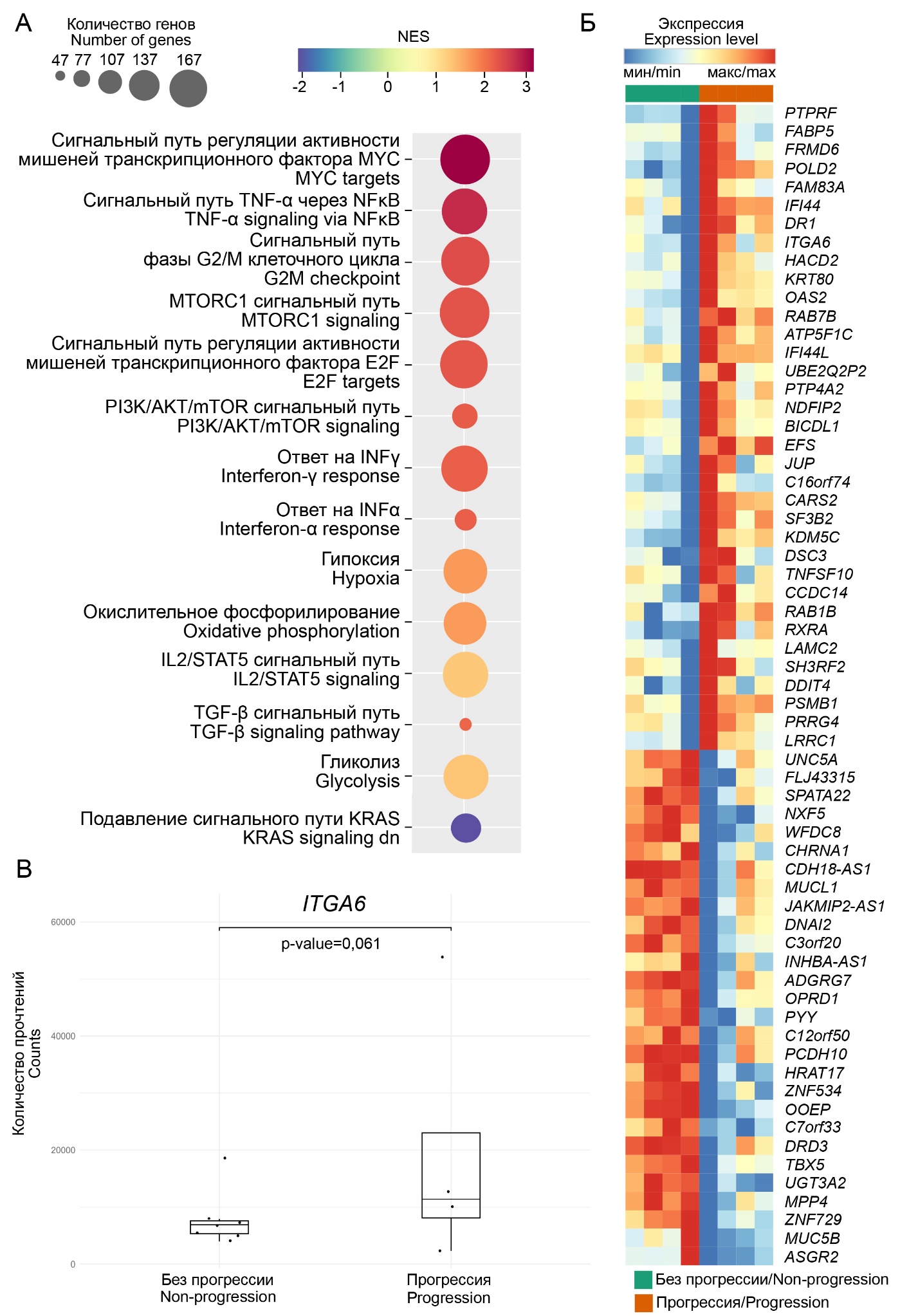
Results. Patients with progressive disease demonstrated activation of oncogenic (TNF-α via NF-κB, PI3K/AKT/mTOR, MTORC1, and KRAS), metabolic (oxidative phosphorylation, glycolysis, and hypoxia), immune (TGF-β, responses to IFNγ and IFNα, and IL2/STAT5), and G2/M cell cycle (through regulation of the E2F and MYC transcription factor targets) signaling pathways. the ITGA6 gene expression level was higher in tumors from patients with cervical cancer progression than in patients without cancer progression.
Conclusion. The progression of stage I-II cervical cancer is driven by a complex interplay of proliferative, metabolic, and immunosuppressive factors, with ITGA6 identified as a potential marker.
ONCOLOGY PRACTICE
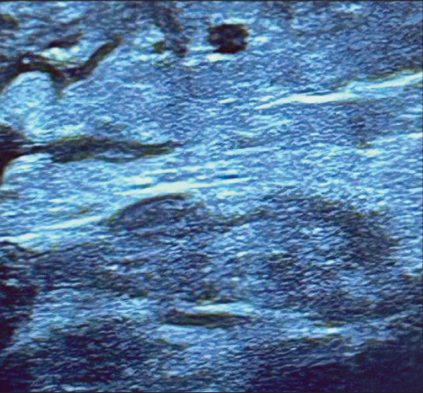
Introduction. Limb lymphedema is a chronic and debilitating disease. it is one of the common postoperative complications following mastectomy with lymph node dissection. lymphedema can be treated conservatively or surgically, including tissue resection and direct or indirect lymphatic drainage. Lymphatic-venous anastomosis (LVA) is an effective surgical treatment for obstructive lymphedema that is not amenable to conservative decongestant therapy. This technique was developed on the basis of supermicrosurgery, a technique of applying anastomoses to vessels with a diameter of 0.3-0.8 mm. Objective: evaluation of the effectiveness and significance of ultrasound examination of lymphatic and venous vessels before the formation of lymphovenous anastomosis.
Material and Methods. The ultrasound diagnostics department, in collaboration with the oncoplastic surgery department of the P.A. Herzen Moscow Oncology Research Institute (Moscow Branch of the National Medical Research Center of Radiology of the Russian Ministry of Health), has accumulated experience examining 135 patients from July 2024 to April 2025. All patients underwent combination treatment for breast cancer. All patients underwent preoperative ultrasound mapping of the superficial subcutaneous veins and lymphatic vessels, as well as lymphography using indocyanine contrast (ICG). During the study, we were specifically interested in the lymphatic and venous vessels.
Results. We achieved 100% coincidence between preoperative markings and the anatomical location of the lymphatic and venous vessels during surgery.
Conclusion. Detection of lymphatic and venous vessels using a high-frequency ultrasound sensor allows for rapid, cost-effective, and time-efficient preoperative mapping, which surgeons can use to reliably guide their lymphatic drainage procedures.
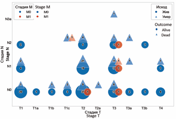
Background. Pancreatoduodenectomy (PD) is one of the most complex abdominal surgeries, associated with significant postoperative morbidity and mortality. advances in modern surgery have reduced postoperative mortality to below 5 % in major centres. However, the frequency of complications remains high, reaching 30-40 %.
Aim of the study: to evaluate the immediate and long-term treatment outcomes of patients with periampullary tumors after pancreatoduodenectomies performed at the Botkin Hospital, Moscow, Russian Federation.
Material and Methods. A retrospective analysis of data from 231 patients with periampullary tumors who underwent pancreatoduodenectomy between January 2015 and December 2025 was conducted in the specialized department of hepatopancreatobiliary surgery at the Botkin Hospital. For evaluating oncological outcomes, general surgical complications were assessed using the Clavien-Dindo classification, and specific complications using the ISGPS classification. A comparative analysis of operative time, blood loss volume, and length of hospital stay was performed. The tumor stage according to the TNM classification and the number of R0/R1 resections were analyzed. Disease-free and overall survival rates, and factors influencing these rates, were evaluated.
Results. Open PD was performed in 180 patients, and robotic PD was performed in 51 patients. The proportion of robotic pancreatoduodenectomies was 22.1 %. The median operative time was 415 minutes, and the mean blood loss volume was 150 ml. R0 resection was achieved in 95.1 % of cases. On average, 15 lymph nodes were removed during surgery. The average length of hospitalization was 18 days. The proportion of severe class IV complications according to Clavien-Dindo was 6.5 %. Class C pancreatic fistula according to the ISGPF classification was diagnosed in 9.5 % of cases postoperatively. The 30-day mortality rate was 5.6 %.
Conclusion. surgical treatment of patients with periampullary tumors is safe and should be performed in high-volume centres. The low level of severe class IV complications according to Clavien-Dindo and 30-day mortality after PD in our centre (6.5 % and 5.6 %, respectively) is similar to rates registered in other major centres and reflects the high level of expertise of the specialized department of the Botkin Hospital in the surgical treatment of patients with periampullary tumors.
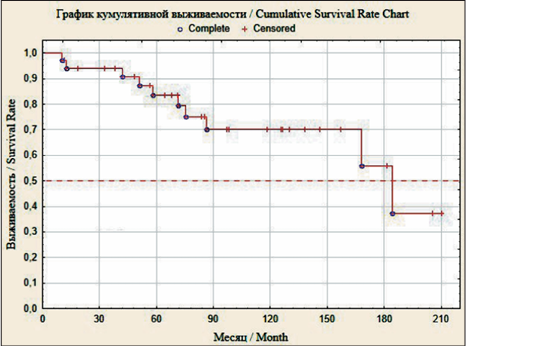
Objective: to analyze clinical data from patients with rectal wall injury after prostatectomy, evaluate long-term surgical and oncologic outcomes, and assess the effectiveness of treatment methods.
Material and Methods. A retrospective analysis of 34 patients with prostate cancer with rectal wall injury was conducted.
Results. Biochemical recurrence was diagnosed in 16 men (47.1 %) with a median of 19.4 months and a median PsA of 0.8 ng/ml. local recurrence was diagnosed in 3 patients (8.8 %) after 3.9 months, 36.0 months and 80.7 months with PsA values of 0.46 ng/ml, 1.275 ng/ml and 5.24 ng/ml, respectively. Generalization of prostate cancer was confirmed in 10 men (29.4 %) with a median of 50.3 months and a median PsA of 18.7 ng/ml. Hormonal resistance of prostate cancer developed in 4 patients (11.8 %). The median follow-up of patients was 85.1 months. One-year mortality was 2.9 %. three patients were lost to follow-up after 12 months, 1.5 years, and 4 years without progression of malignant neoplasms. ten deaths were confirmed from malignant neoplasms (n=6), circulatory diseases (n=3), and unlawful acts committed against the patient (n=1). at the time of follow-up, 21 patients (67.7 %) were alive, of whom 12 (57.1 %) showed no signs of prostate cancer progression.
Conclusion. in case of intraoperative and perioperative disruption of the integrity of the rectal wall after prostatectomy, restoration of the rectal defect or long-term carriage of urinary and/or intestinal stomas, possibly lifelong, does not affect the course of the tumor process and is not a direct cause of a reduction in life expectancy in men.
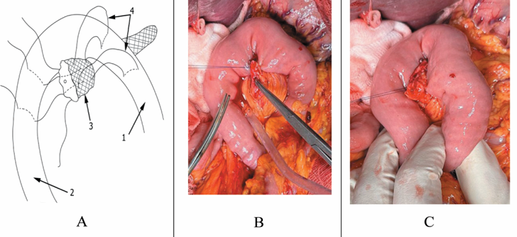
Introduction. In this paper we present a novel sleeve-shaped pancreaticojejunostomy (NSSP) technique developed for pancreaticoduodenectomies in case of soft pancreatic parenchyma.
Material and Methods. The efficacy of NSSP was compared to “duct-to-mucosa” (DTMA), invaginational (IA), and catheter-guided (CGA) types of pancreatic anastomoses. A total of 173 pancreaticoduodenectomies were analyzed.
Results. The rate of clinically relevant postoperative pancreatic fistulas (CR-POPF) was 6.6 % in the group of “duct-to-mucosa” anastomoses, 16.0 % in the group of invaginational anastomoses, and 22.2 % in the group of catheter-guided ones. CR-POPF have not been registered in the group of NSSP. The risk of CR-POPF after application of NSSP was significantly lower compared to the group of IA (OR 3.44 [2.63-11.11]), the group of CGA (OR 5.54 [3.41-8.22]) and did not differ from the group of DTMA (OR 1.19 [0.65-2.38]).
Conclusion. The novel sleeve-shaped pancreaticojejunostomy has proved to be effective and in a short series of surgeries demonstrated the lowest rate of CR-POPF compared to other known types of anastomoses.
REVIEWS
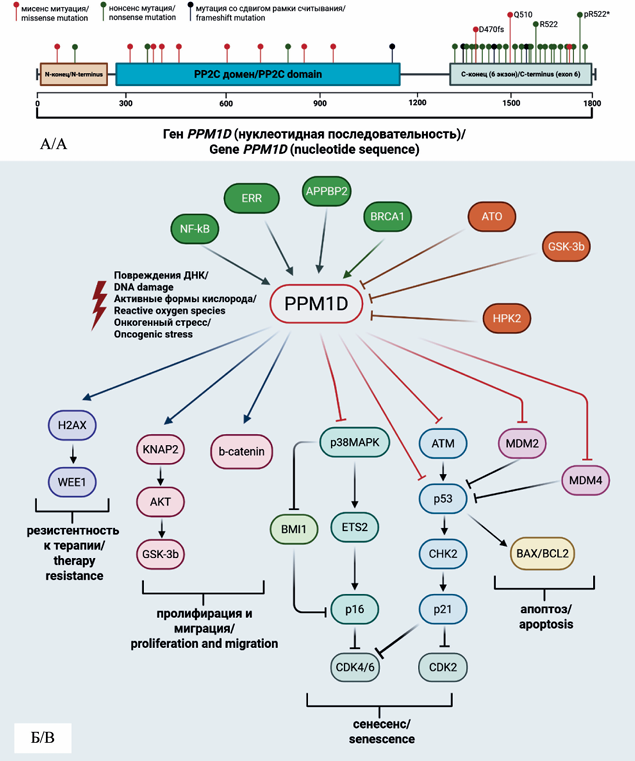
Objective: to summarize the accumulated knowledge on the role of PPM1D phosphatase in clonal hematopoiesis and the pathogenesis of hematological malignancies, particularly in acute myeloid leukemia, as well as to evaluate the feasibility of therapeutic targeting of PPM1D using specific inhibitors.
Materials and Methods. A search for relevant sources was conducted in the Web of Science, PubMed, and Scopus databases. Publications were selected based on the relevance of the studies and the pertinence of their subject matter to the topic of the review, the literature search was performed using the following key terms: “mutations of PPM1D", “PPM1D and cancer”, “clonal hematopoiesis”, “PPM1D and clonal hematopoiesis”, “PPM1D gene”, “WIP1 phosphatase”, “clonal hematopoiesis of indeterminate potential”, “therapy-related AML”, “p53 signaling pathway”, “cell cycle regulation”, “targeted cancer therapy”, “hematologic malignancies”, “truncating mutations” and “inhibitors of PPM1D”. A total of 142 sources were analyzed, of which 63 were included in the review.
Results. Clonal hematopoiesis of indeterminate potential (CHIP) is characterized as a condition involving somatic mutations in driver genes of myeloid neoplasms at a variant allele frequency of ≥2 % in blood cells of individuals without hematological disorders. The prevalence of CHIP exceeds 10 % in individuals over 60 years of age and reaches 25 % in cancer patients. Antineoplastic therapy promotes the selection of hematopoietic stem cell clones harboring mutations, including mutations in the PPM1D gene, which substantially increases the risk of developing therapy-related myeloid neoplasms (t-MN). PPM1D phosphatase is a negative regulator of p53 and numerous cell death pathways. Its overexpression is detected in various types of solid tumors (ovarian cancer, breast cancer, and others), while its mutations are identified in 1-2 % of patients with de novo myeloid neoplasms and in 10-20 % of patients with t-MN. Recent studies demonstrate the predominant role of PPM1D mutations in age-associated clonal hematopoiesis, in the context of telomere shortening, and in the presence of germline DNA repair mutations, thereby underscoring the pivotal significance of this phosphatase in the pathogenesis of myeloid disorders.
Conclusion. PPM1D represents a promising therapeutic target for the treatment of AML owing to its key role in the regulation of multiple cell death pathways and the cellular stress response. PPM1D inhibitors may serve as a foundation for the development of combination therapy regimens, particularly for elderly patients and those with acquired resistance to antineoplastic agents.
Objective. This review summarizes the current state of development of oncolytic virus-based therapeutics, including already approved agents, candidates in clinical and preclinical trials, and emerging trends in oncolytic virotherapy as of 2025.
Material and Methods. A systematic literature search was performed in Web of Science, Scopus, PubMed and the Russian Science Citation Index (RSCI) covering the period from 1990 to 2025. The review includes peer-reviewed publications indexed in Web of Science and Scopus as well as clinical trial reports (phases I–III ).
Results. By 2025, four oncolytic virus products have been approved for clinical use: Talimogene laherparepvec (T-VEC, Imlygic®), Teserpaturev (G47Δ, Delytact®), Oncorine (H101), and Rigvir. Their therapeutic effect is mediated by selective replication in tumor cells followed by cell lysis, combined with the induction of innate and adaptive immune responses. Ongoing clinical trials investigate additional candidates, including adenovirus-based therapies (CG0070, DN X-2401, ONCOS-102), vaccinia virus (Pexa-Vec), and modified herpes simplex viruses (Seprehvir). Novel research efforts also focus on Newcastle disease virus (ND V; strains MTH-68/H, PV701, ND V-HUJ), measles virus, Sendai virus, vesicular stomatitis virus, and coxsackievirus A21. Advances in genetic engineering, such as deletion of virulence genes, insertion of immune-modulatory transgenes, and the use of genome editing tools have enhanced the selectivity and safety of viral constructs. In addition, the exploration of wild-type strains as potential oncolytic agents is emerging as a promising field.
Conclusion. Oncolytic virotherapy is transitioning into an established component of cancer treatment. Major trends include the personalization of therapy, integration with immune checkpoint inhibitors and cell-based strategies, and the application of advanced genome editing technologies. The investigation of wild-type viral strains remains an important future direction for identifying novel oncolytic agents.
CASE REPORTS
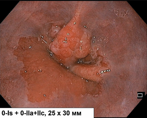
Background. Early esophagogastric junction cancer with submucosal invasion (pT1b) has traditionally been an indication for radical esophagectomy due to the risk of lymphatic metastasis. However, accumulating evidence points to heterogeneity in the biological behavior of these tumors: in patients with a favorable histological profile (low-grade differentiation, absence of lymphovascular and perineural invasion), the rate of metastasis may be comparable to postoperative mortality associated with esophagectomy. A personalized organ‑preserving approach including endoscopic submucosal dissection (ESD) and intraoperative indocyanine green (ICG) navigation of sentinel lymph nodes may potentially avoid debilitating surgery in selected patients. Nevertheless, the safety and efficacy of this strategy for deep submucosal invasion (sm2, depth >500 μm) remain controversial.
Case presentation. A 63‑year‑old patient with early esophagogastric junction cancer (cT1aN0M0, Siewert type II ) underwent combined modality treatment including laparoscopic ICG navigation with removal of two sentinel lymph nodes along the lesser curvature of the stomach (frozen‑section analysis showed no metastases) and simultaneous endoscopic submucosal dissection (ESD). Final histopathological examination of the resected specimen revealed a well‑differentiated adenocarcinoma pT1b sm2 (>500 μm) with no lymphovascular or perineural invasion, a negative vertical margin, but scattered tumor cells in the coagulated horizontal margin. Two months later, residual tumor was diagnosed and repeat ESD achieved R0 resection. Seventeen months after the primary procedure, a solitary lymphogenous metastasis was detected along the left gastric artery. Laparoscopic selective lymphadenectomy and adjuvant polychemotherapy (8 cycles of XELOX) were performed. No signs of recurrence were observed 4.5 years after repeat ESD and 3 years after lymphadenectomy.
Conclusion. This case report demonstrates that a personalized organ‑preserving approach (ESD with ICG‑guided sentinel lymph node navigation) can be implemented in carefully selected patients with early high‑risk esophagogastric junction cancer (pT1b sm2) and favorable histological characteristics (G1–2, L0, V0). Strict multimodal surveillance enables timely detection of oligometastatic progression and selective lymphadenectomy, thereby avoiding esophagectomy and preserving quality of life. Further data accumulation is needed to refine patient selection criteria and optimize follow‑up protocols.
Background. According to the International Morphological Classification of Diseases for Oncology, lung carcinoids are classified as malignant epithelial neoplasms. However, carcinoid tumors are rare: less than 6 cases per 100,000 people annually. A typical carcinoid often manifests as a small (less than 3 cm), slowly growing peripheral lung tumor that rarely metastasizes (less than 1 % of cases). However, even with small tumor sizes, approximately 3 % of carcinoids are hormone-producing and can present with paraneoplastic syndromes. The primary treatment for carcinoid tumors is surgery; however, the volume of lung resection, particularly for stage IA peripheral carcinoids, is a subject of intense debate in the current literature.
The objective of this study was to present a case of successful reoperation involving a right upper lobectomy using a minimally invasive approach for local recurrence of an ACTH-producing lung carcinoid. The postoperative period was uneventful and clinical manifestations resolved completely.
Case description. Patient S., 32, with a confirmed typical carcinoid pT1a cN0M0 with pronounced clinical manifestations of increased ACTH production, experienced a recurrence of symptoms 15 months after an atypical resection of the right upper lobe, along with local tumor recurrence in the form of intrapulmonary lymph node involvement. This situation necessitated a repeat radical surgery involving a thoracoscopic extended right upper lobectomy 2 years after the initial surgery. The postoperative period was uneventful. A follow-up examination after 3 months revealed complete regression of symptoms associated with increased ACTH production.
Conclusion. Despite the low prevalence of carcinoid tumors and their low malignancy potential, surgical interventions should adhere to the same oncological principles as for other forms of lung cancer, especially if the tumor is hormonally active. Furthermore, the clinical manifestations of a hormone-producing tumor can allow for early detection of local recurrence or disease progression. When choosing the extent of surgery in patients with carcinoid tumors, it is important to consider not only the tumor size and location but also its hormonal activity. In these cases, radical anatomical lung resection with lymph node dissection is advisable for adequate staging and improved long-term treatment outcomes.
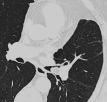
Benign neoplasms of the tracheobronchial tree are rare, accounting for approximately 7-10 % of all primary lung tumors. Endobronchial lipoma is an extremely uncommon benign tumor, comprising approximately 0.5 % of all bronchial tumors. These benign tumors typically have a rounded shape, a distinct capsule, and an elastic consistency. Morphologically, lipomas appear as mature adipose tissue separated by connective tissue septa. Intrabronchial location within the tracheobronchial tree is the most common; this type of tumor growth is observed 4-5 times more often than perior exobronchial lesions. The absence of pathognomonic symptoms and clinical manifestations, coupled with the slow growth of the tumor, often leads to late diagnosis. The primary goal in treating endobronchial lipomas is to eliminate bronchial obstruction and prevent irreversible changes to lung tissue. For large tumors obstructing the bronchial lumen or the presence of significant secondary changes to the lung tissue, endoscopic removal is not advisable. Surgical treatment, including segmentectomy or lobectomy, is the optimal option. Here we present a case of successful combined diagnostic approaches for rare endobronchial lipoma.
Case presentation. A 79-year-old man presented with benign tumor in the left bronchial tree, with minimal respiratory symptoms. A chest computed tomography revealed a fat-density lesion within the lumen of the left lower lobe bronchus with partial obstruction of the bronchial lumen. Bronchoscopy demonstrated a mass lesion extending into the lower third of the left main bronchus.
Conclusion. The presented clinical case demonstrates the challenges associated with the timely diagnosis of endobronchial lipomas and highlights the importance of the multidisciplinary approach utilizing imaging, endoscopic, and histopathological examination for diagnostic verification and treatment planning.
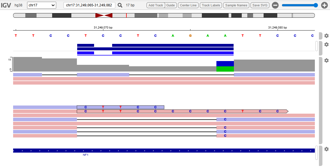
Background. Neurofibromatosis type 1 is a common hereditary autosomal dominant disorder caused by pathogenic genetic variants in the NF1 gene located on chromosome 17q11.2. The disease is characterized by high allelic heterogeneity and the absence of clear genotypic correlations, with the exception of large deletions associated with a more severe phenotype. Clinically, neurofibromatosis is characterized by the presence of multiple (≥6) ‟café-au-lait” spots, neurofibromas of any type or plexiform neurofibromas, freckles in the axillary or inguinal areas, hamartomatous Lisch nodules of the iris, optic glioma, and bone dysplasia. Therefore, the description of each new genetic variant is essential for expanding the spectrum of known variants and improving molecular diagnostics.
Case Report. The article describes a clinical and molecular genetic study of a 10-year-old boy. Since early childhood, the patient has had multiple “café-au-lait” spots, speech impairment, grade 1 hypotrophy, rickets, sequelae of perinatal CNS damage, myotonic syndrome, and a delay in motor development. Magnetic resonance imaging of the brain revealed signs of focal damage to both hemispheres, the cerebellar vermis, and the left subcortical nuclei, and a glioma of the right optic nerve was detected. Neurofibromatosis type 1 was diagnosed based on clinical criteria. Whole-genome DNA sequencing was performed, followed by bioinformatics analysis and confirmation of the results by direct Sanger sequencing. A previously undescribed complex, likely pathogenic variant NM_001042492.3:c.40 60_4068delinsC of the NF1 gene was identified in the heterozygous state. This variant leads to a reading frameshift and premature translation termination after the synthesis of 23 amino acids (p.Ser1354Leufs*23). This variant is not present in available population genetic variant databases.
Conclusion. Thus, whole-genome sequencing identified a new pathogenic variant in the NF1 gene associated with the development of neurofibromatosis type 1. The obtained data expand the range of variants for this pathology and can be used in medical genetic counseling, as well as in algorithms for molecular genetic diagnostics of patients with suspected neurofibromatosis type 1.
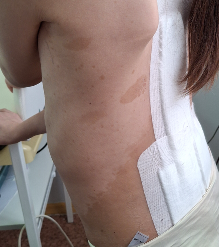
Background. Neurofibromatosis type 1 (NF1) is an autosomal dominant tumor syndrome characterized by marked polymorphism of clinical manifestations. There is evidence of genotypic correlations in NF1 with more pronounced manifestations of the disease with certain mutations in the NF1 gene. therefore, it is important to describe patients with a specific mutation and a severe NF1 phenotype.
Purpose of the study: to describe the genetic and clinical features of NF1 and its treatment tactics in the patient with severe manifestations of the disease and a unique mutation in the NF1 gene.
Material and Methods. A ten-year-old girl with a sporadic case of NF1 was examined, X-ray examination was performed, a blood sample was taken with DNA extraction and sanger sequencing of the NF1 gene.
Results. The patient was identified to have a unique pathogenic variant c.240_241del(p.Y80fs) in the NF1 gene, and the following clinical manifestations of NF1: retrocerebellar brain cyst, femur plexiform neurofibroma, grade 3 scoliosis, and femur fibrous dysplasia. successful surgical correction of the scoliosis was performed. targeted therapy with selumetinib was prescribed for femur plexiform neurofibroma treatment.
Conclusion. the identified NF1 variant: c.240_241del(p.Y80fs) has not previously been described in the scientific literature and is not included in the ClinVar database. the clinical manifestations of NF1, characterized by severe combined lesions, have been described. treatment of such cases of NF1 requires a combination of targeted therapy and high-tech surgery.
ANNIVERSANES


ISSN 2312-3168 (Online)